-
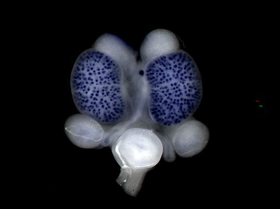
In situ staining of Lhx1 on an embryonic day 15.5 urogenital system (Image by Lisa Rutledge) -

An embryonic day 15.5 kidney stained with PECAM (red, blood vessels), Tuj1 (blue, neurons) and nephrin (yellow, podocytes). The staining procedure was iDISCO. (Image by Riana Parvez and Seth Ruffins) -
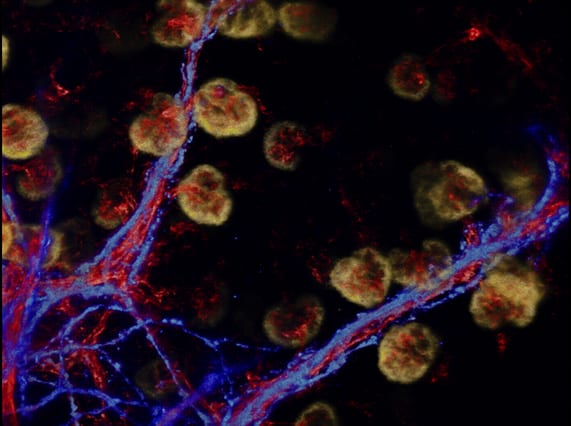
An embryonic day 15.5 kidney stained with PECAM (red, blood vessels), Tuj1 (blue, neurons) and nephrin (yellow, podocytes). The staining procedure was iDISCO. (Image by Riana Parvez and Seth Ruffins) -
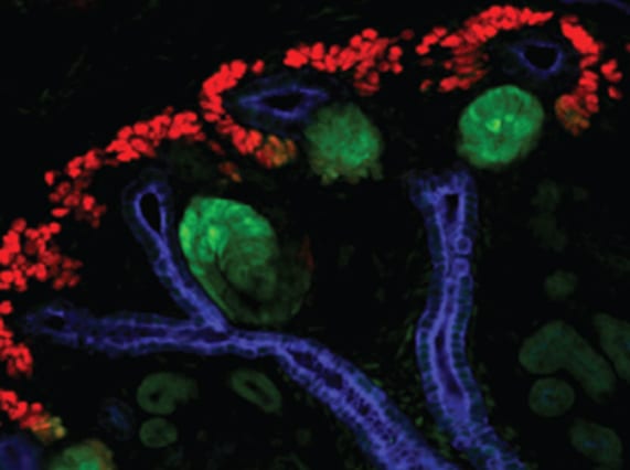
Embryonic kidney showing nephron progenitors (red), developing nephrons (green) and the collecting duct (blue) (Image by Lori O'Brien) -

Embryonic kidney highlighting the nephron progenitors (green) and collecting duct (red) (Image by Lori O'Brien)
